PARKIN Autopsy Reports And PRKN-Related Clinical Phenotype Notes
Regional Neuronal Loss And Gliosis
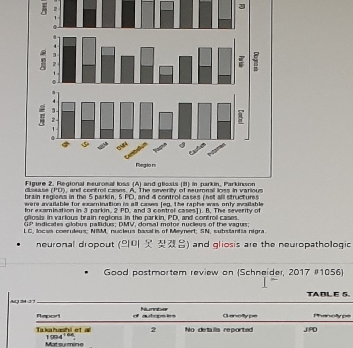
Figure 2 material continues from 20240722_181943.
Caption text:
Figure 2. Regional neuronal loss (A) and gliosis (B) in parkin, Parkinson
disease (PD), and control cases. A, The severity of neuronal loss in various
brain regions in the 5 parkin, 5 PD, and 4 control cases (not all structures
were available for examination in all cases [eg, the raphe was only available
for examination in 3 parkin, 2 PD, and 3 control cases]). B, The severity of
gliosis in various brain regions in the parkin, PD, and control cases.
GP indicates globus pallidus; DMV, dorsal motor nucleus of the vagus;
LC, locus coeruleus; NBM, nucleus basalis of Meynert; SN, substantia nigra.
Note:
neuronal dropout (의미 못 찾겠음) and gliosis are the neuropathologic findings
Movement Disorders, Vol. 28, No. 6, 2013
Schneider 2017 PARKIN Autopsy Reports
Note above the table:
Good postmortem review on (Schneider, 2017 #1056)Table title:
TABLE 5. Summary of PARKIN autopsy reportsTable headers:
Report
Number of autopsies
Genotype
Phenotype
Pattern of neuronal loss
LB, LN pathology
LB distribution - Braak stage
Tau pathology - NFT stage
Other inclusions
Selected row-level anchors:
| Report | Number of autopsies | Genotype | Phenotype | Pattern / pathology fragments |
|---|---|---|---|---|
Takahashi et al 1994 | 2 | No details reported | JPD | LC>SNpc; n.d. in tau/NFT column |
Matsumine et al 1997; Yamamura et al 1998 | 1 | Homozygous del between exon 3 and 7 | EOPD | SNpc>LC |
Mori et al 1998 | 1 | Homozygous exon 4 del | EOPD | SNpc>LC; tau/NFT 3; Thorn-shaped astrocytes |
Hayashi et al 2000; van de Warrenburg et al 2001 | 1; 1 | Homozygous exon 4 del; Compound heterozygous exon 3 del/exon 6 transversion | EOPD | SNpc>SNpr, LC; SNpc>LC; Sparse; Diffuse in the caudate nucleus, putamen, subthalamic nucleus, and SN; Thorn-shaped astrocytes |
Farrer et al 2001 | 1 | Compound heterozygous exon 7 R275W/exon 3 del | EOPD, writer's cramp | SNpc, LC; +; Braak 4 |
Mori et al 1998; Gouider-Khouja et al 2003; Sasaki et al 2004/2008 | 1; 1; 1 | Compound heterozygous exon 6 del/exon 7 del; Homozygous exon 2 del; Homozygous exon 3 del | EOPD | includes SNpc>LC, SNpc, SNpr>LC, Basophilic LB-like in PPN, and Eosinophilic LB in anterior horn cells |
Pramstaller et al 2005; Orimo et al 2005 | 1; 3 | Compound heterozygous exon 7 del and 1072T del; Homozygous exon 4 del | PD; EOPD | SNpc, LC; n.d.; Numerous TH-immunoreactive nerve fibers in the epicardium |
Miyakawa et al 2013 | 1 | Homozygous exon 2-4 del | Late-onset PD | SNpc, LC, severe; + , fairly widespread; Braak 4; Yes; Eosinophilic inclusions with HE, TH, and phosphorylated neurofilament in epicardium |
Doherty et al 2013 | 5 | R275W/del exon 6; R275W/Pro113fs; R275W/G430W; G430D/Pro113fs; R275W/del exon 6 | EOPD (2), IPD (3) | Moderate to severe in SNpc, mild to moderate in LC; SNpc>LC in all; + (in 2), - (in 3); Absent (in 2) or only mild (in 3 cases); TDP-43-positive inclusions absent |
Cornejo-Olivas et al 2015 | 1 | Compound heterozygous intron 5 splice site mutation (IVS5-1G>A)/exon 7 del | JPD | SNpc; n.d.; TH immunopositive fibers in striatum |
Morales et al 2002; Ruffmann et al 2012; Sharp et al 2014 | 1; 1; 1 | Heterozygous C212Y mutation; Heterozygous R275W mutation; Heterozygous exon 3-4 del | PSP; IPD, onset 62 y; EOPD | SNpc/pr, striatum, GP, nbM, STN, thalamus; Severe in SN and LC; LB/LN +; Braak 6; Pre-tangles in subiculum, transentorhinal, and entorhinal cortex; Widespread cortical deposition of BAP |
Footnotes/abbreviation text:
Patients, that is, homozygous and compound heterozygous mutation carriers, are shaded in light gray.
Single mutation carriers are shown in white cells.
del, deletion; SNpc, substantia nigra pars compacta; SNpr, substantia nigra pars reticulata;
GP, globus pallidus; LB, Lewy body; LN, Lewy neurites; TH, tyrosine hydroxylase;
HE, haematoxylin and eosin stain; BAPs, beta-amyloid plaques; EOPD, early-onset PD;
JPD, juvenile-onset PD; n.d., no data.
*The pattern of pathology did not conform well to the Braak PD staging scheme as the density of brain stem LBs did not show the expected increase when LB pathology extended beyond the brain stem.
Immunohistochemical study of heart tissues, not brain.
Clinical Phenotype Notes
Notes:
clinical phenotype

- juvenile Parkinsonism: AR, early disease onset (mean age of onset: without LBs,
  postmortem, 27 years) and a marked response to levodopa (Khan et al., 2003);
- prominent lower extremity hyperkinetic movements (i.e., foot dystonia, leg tremor)
  and relative lack of nonmotor features
- long term outcome
  - Long-term survival of patients with PRKN-related PD is usually favorable.
    Although they have a low age at disease onset, most of them have a long disease duration

- Dx
  - The diagnosis of parkin type of early-onset Parkinson disease can only be confirmed
    when pathogenic variants are identified on both alleles of PARK2
    9IE HOMOZ OR COMPOUND HETEROZ) 2017 Ivashynka
Comparison table/note fragments:
familial
sporadic

clinically indistinguishable from idiopathic PD (2017 Ivashynka).

- median age at onset is 31 years (range: 3-81 y) (Brüggemann N, 2020 #779)
- Lower-limb dystonia in 65% (may be a presenting sign or may occur during disease progression),
- Well-preserved sense of smell
- Hyperreflexia (50%)
- Slowly progressive: (Alcalay, 2014 #1034, N=21, cross sectional, but they perform better on UPDRS III)
- [Pyatigorskaya et al 2015].
- [Nonmotor Sx]
- (Yoritaka, 2011 #1033): js: olfactory dysfunction is less common than Spd
- (Alcalay, 2014 #1034, N=21, cross sectional, table 1.
  (authors say, dementia diagnosis was made by clinical consensus, that's why 9.5% despite zero 1/2 on CDR)
The left edge of the comparison table is partly cut off; OCR suggests fragments including familial, juvenile Parkinson's, EOPD, 10% of total PD, and (Camargos, 2009 #689).
Alcalay 2014 EOPD PARKIN Cognitive/Demographic Table
Table title:
Table 1
Clinical and demographic characteristics of EOPD probands with and without PARKIN compound heterozygote/homozygote mutations
Table transcription:
| Characteristic | Non-Carriers N=23 | Two mutations (compound heterozygotes/homozygotes) N=21 | P value |
|---|---|---|---|
| Mean Age, years (SD) | 61.5 (6.4) | 53.1 (11.5) | 0.004 |
| Mean Age at onset, years (SD) | 40.2 (4.0) | 26.6 (10.0) | <0.001 |
| Mean Disease duration, years (SD) | 21.3 (4.2) | 26.5 (9.7) | 0.023 |
| Mean Education, years (SD) | 15.7 (4.2) | 13.5 (2.8) | 0.052 |
| Neuropsychological testing in Spanish (Number of subjects) | 13.0% (3) | 23.8% (5) | 0.448 |
| Mean UPDRS-III (SD) | 27.8 (10.1) | 21.0 (7.0) | 0.015 |
| Mean Levodopa equivalent daily dose, mg (SD) | 811 (366) | 650 (530) | 0.252 |
| Ethnicity (Caucasian/Hispanic/Other %) | 73.9%/21.7%/4.3% | 61.9%/38.1%/0.0% | 0.343 |
| Gender (% Female) | 9 (39.1%) | 12 (57.1%) | 0.365 |
| Mean MMSE | 27.9 (2.0) | 29.2 (0.9) | 0.010 |
| CDR 0 | 40.9% (9) | 76.2% (16) | 0.003 |
| CDR 0.5 | 9.1% (2) | 23.8% (5) | |
| CDR 1 | 40.9% (9) | 0.0% | |
| CDR 2 | 9.1% (2) | 0.0% | |
| Clinical diagnosis - Normal | 21.7% (5) | 28.6% (6) | 0.018 |
| Clinical diagnosis - Mild cognitive impairment | 30.4% (7) | 61.9% (13) | |
| Clinical diagnosis - Dementia | 47.8% (11) | 9.5% (2) | |
| Mean Psychomotor speed, Z-score (SD) | -0.58 (1.1) | -0.11 (0.74) | 0.114 |
| Mean Attention, Z-score (SD) | -0.64 (0.95) | 0.00 (0.83) | 0.027 |
| Mean Memory, Z-score (SD) | -0.51 (0.91) | -0.14 (0.79) | 0.162 |
| Mean Visuospatial function, Z-score (SD) | -0.44 (1.3) | 0.10 (0.59) | 0.080 |
| Mean Executive function, Z-score (SD) | -0.53 (0.90) | -0.07 (0.63) | 0.057 |
Footnotes:
Four non-carrier participants completed only portions of the neuropsychological examination.
one non-carrier did not receive a CDR score
Hattori note:
Hattori:
rigidity 적음,
freezing gait 로 인해 잘 넘어짐.
Slowness 있음.
Uncertain Spans
page_labelis taken from the visible Word statusbar:Page 98 of 104. The same Word page spans multiple photos.- The statusbar/body-overlap
Longitudinalnote is visible only through the statusbar crop. BG maturation (20 y) 이후에 develop,DBS response is good,Gut is important, (vagus n stimulation), and(Sun, 2021 #1647)fragments sit near the bottom/statusbar overlap.- In the autopsy table, report citation/year, genotype, phenotype, brain-region abbreviation, LB/LN status, Braak/NFT stage, and “Other inclusions” cells are small-text transcriptions.
9IE HOMOZ OR COMPOUND HETEROZ)in the Dx note is visibly awkward and may be a cropped/corrupted insertion.Brüggemann N, 2020 #779,(Camargos, 2009 #689),(Yoritaka, 2011 #1033),(Alcalay, 2014 #1034), and[Pyatigorskaya et al 2015]citation strings should be verified.- Cognitive table values
53.1,26.6,26.5,21.0,29.2,76.2%,23.8%,61.9%,9.5%, all p-values, and all Z-scores are small-text table values. - The CDR and clinical-diagnosis p-values appear as block-level p-values rather than per-subrow p-values; the Markdown table leaves lower subrow p-value cells blank accordingly.
- The Hattori note is at the bottom edge and may continue on the next photo.