PRKN Heterozygous Carrier Frequency, EOPD Risk, And Neuropathology
Heterozygous PRKN Carrier Notes
Table2) pathogenic SNV & CNV heterozygous carrier 가, 약 2000 명 이상의 PD 환자중 1.2%,
약 2000 명 이상의 control 에서 1.8% 로 차이부 (오히려 PD 에서 적음, JS: This tells us that
heteroz PRKN mutation is not a risk factor for LOPD 왜냐면 LOPD 환자가 majority
였으니까,)

Pathogenic SNV & CNS is 14% of total (SNV & CNV) (1.52/10.7)

Pathogenic Parkin variant should mean the variants that show reduction of either parkin
function or parkin expression (applied in 2019 Yi). Target? Controversial, so is there any
means to address this?
CNS in the visible note may be an OCR/typing issue for CNV; it is left as visible/uncertain text rather than silently corrected.
EOPD-Only Carrier Analysis
Note:
[EOPD only]
Is it a risk factor for EOPD? -> (table S5) no association (p>0.05), but trend (OR=~1.5)
Pathogenic SNV & CNS is ~20% of total (SNV & CNV)
Table transcription:
| analysis in participants below 50 | Carriers in patients, No. (%) | Carriers in controls, No. (%) | SKAT-O |
|---|---|---|---|
| SNV | 50 (12.5) | 276 (9.38) | 0.507 |
| CNV | 7 (1.75) | 29 (0.982) | 0.594 |
| SNV & CNV | 55 (13.7) | 293 (9.9) | 0.584 |
| Patho SNV | 5 (1.25) | 23 (0.778) | 0.814 |
| Patho SNV & CNV | 12 (2.99) | 52 (1.76) | 0.765 |
| No Benign SNV | 34 (8.48) | 186 (6.3) | 0.507 |
| No Benign SNV & CNV | 40 (9.97) | 203 (6.87) | 0.589 |
Additional visible row notes:
Early vs late not specified

Comp Bio 보자!
PD=1,272
Prodromal=57
HC=676

{Huttenlocher, 2015 #1924}
1415 PD patients and 40 474 controls ≥65 y
OR=1.67. 4.55% in total 인듯 (ie PD+ non PD), Arg275rp 가 mocst common.
Population Estimates And Penetrance
Source label:
Takeda internal| Group (age ≥ 18) | Biallelic PRKN mutations in general population | Biallelic PRKN mutations with symptomatic PD | Monoallelic PRKN mutations with symptomatic PD |
|---|---|---|---|
| United States and Europe | 23,000-70,000 | 1,622-13,000 | 33,000-45,000 |
| United States | 10,000-30,000 | 930-7,400 | 19,000-26,000 |
| Europe (EU + UK) | 13,000-40,000 | 692-5,500 | 14,000-19,000 |
| Japan | 40-107 | NA | NA |
| China | 420-1,100 | NA | NA |
Penetrance note:
Penetrance of Heterozygus parkn mutation

{Castelo Rueda, 2021 #1923}
Mean age: 45
Penetrance is (2.44-) 7.5% (table s1,
PDS11, ie PD medication)
Neuropathology Notes
[Poulopoulos et al 2012]. The most prominent and most common feature was the finding of neuronal loss in pigmented nuclei of the brain stem. Unlike Parkinson
disease of other etiologies, the neuronal loss was greater in SNpc than in LC (see Parkinson Disease Overview). Typical alpha-syn-containing Lewy bodies were identified
in only two affected individuals, whereas one affected individual had basophilic Lewy body-like pathology of the pedunculopontine nucleus. Tau-containing
neurofibrillary tangles were observed in two affected individuals. In summary, the spectrum of postmortem findings is broad and thus reminiscent of the situation in
LRRK2 Parkinson disease [Kasten et al 2018].

{Doherty, 2013 #778}: review논문 아니지만, 다른 postmortem study 결과 많이 게재함.
LB/a-syn
however, there is a distinct lack of LB formation

No juvenile-onset cases have ever been reported with LBs postmortem.22,23 Movement Disorders, Vol. 28, No. 6, 2013

In a relatively large series of patients with PRKN-related PD and a review of the literature, together with autopsy reports of 13 patients with PRKN-related PD, only 3 out of 13 had
any LBs. (Aasly, 2020 #781)

{Seike, 2021 #1271} 3 out of 8 patients show LB (한명: brain-stem predominant, 두번째환자: limbic subtypes, 세번째환자: lc, DORSOVAGAL NUCLEUS, BUT NOT IN snPC)
Neuronal Loss
{Doherty, 2013 #778}:

of course SN,

Mild neuronal loss in the locus coeruleus and dorsal motor nucleus of the vagus (in the medulla) (mild in 2 of 3 cases examined) and
the cerebellum (js: cerebrum 아님!) (examined at the level of the dentate nucleus and the superior cerebellar peduncle), which showed
mild Purkinje cell loss with empty baskets,

but not in the nucleus basalis of Meynert, raphe nucleus, or other brain regions

{Seike, 2021 #1271} variable degree of neuronal loss in SN & LC
Gliosis
Mild to moderate gliosis accompanied the neuronal loss previously described,

also evidence of gliosis (in the absence of detectable neuronal loss) in the raphe nucleus, NBM, striatum, globus pallidus, dentate nucleus,
amygdala, hippocampus, and cerebral cortices (Figure 2B).

{Seike, 2021 #1271} mild to moderate gliosis in SN & LC
Regional Neuronal Loss And Gliosis Figure
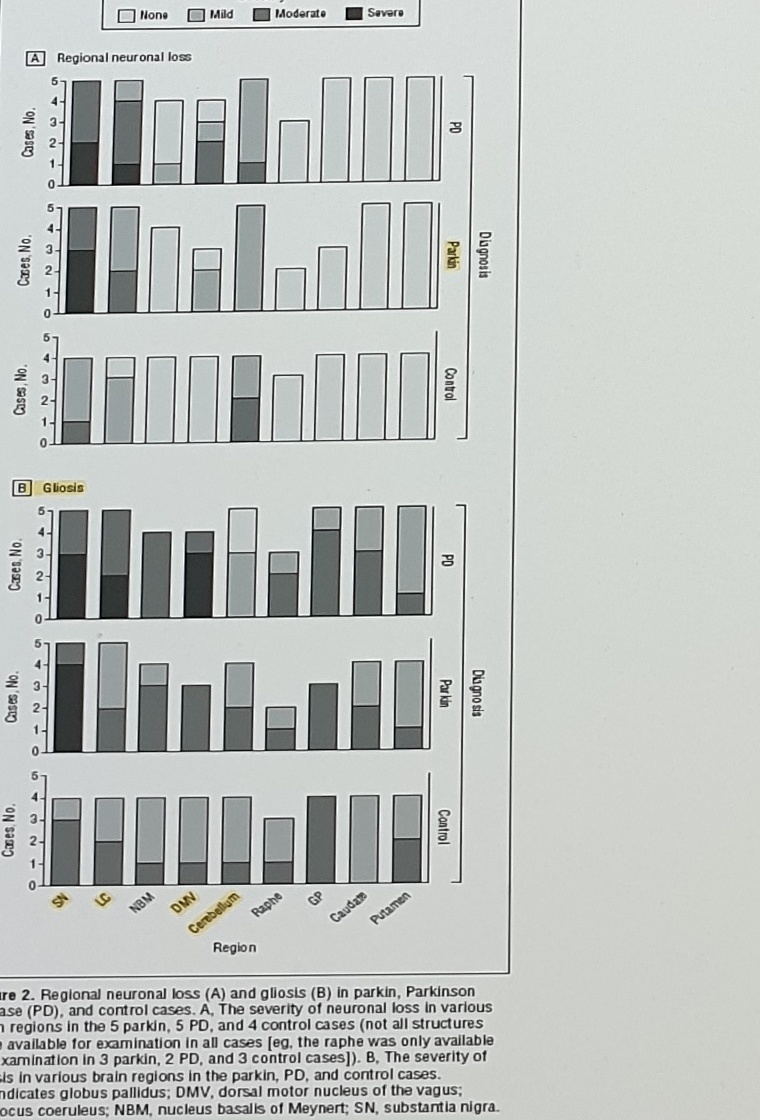
Figure/caption text:
Figure 2. Regional neuronal loss (A) and gliosis (B) in parkin, Parkinson
disease (PD), and control cases. A, The severity of neuronal loss in various
brain regions in the 5 parkin, 5 PD, and 4 control cases (not all structures
were available for examination in all cases [eg, the raphe was only available
for examination in 3 parkin, 2 PD, and 3 control cases]). B, The severity of
gliosis in various brain regions in the parkin, PD, and control cases.
GP indicates globus pallidus; DMV, dorsal motor nucleus of the vagus;
LC, locus coeruleus; NBM, nucleus basalis of Meynert; SN, substantia nigra.
Note below the caption:
neuronal dropout (의미 못 찾겠음) and gliosis are the neuropathologic findings Movement Disorders, Vol. 28, No. 6, 2013Schneider 2017 PARKIN Autopsy Reports
Note:
Good postmortem review on (Schneider, 2017 #1056)Table title:
TABLE 5. Summary of PARKIN autopsy reportsHeader and first row. The table continues beyond the bottom of this photo.
| Report | Number of autopsies | Genotype | Phenotype | Pattern of neuronal loss | LB, LN pathology | LB distribution - Braak stage | Tau pathology - NFT stage | Other inclusions |
|---|---|---|---|---|---|---|---|---|
| Takahashi et al 1994 | 2 | No details reported | JPD | LC>SNpc | - | - | n.d. | - |
Uncertain Spans
Page 98 of 104is from the visible Word statusbar and conflicts with earlier synthesized pages that used a different denominator.CNSinPathogenic SNV & CNSappears in OCR/visible text but probably refers toCNV; it is not normalized here.OR=1.67. 4.55% in total 인듯has ambiguous punctuation; it may represent separate values rather than one sentence.Arg275rp 가 mocst commonis left close to the visible text. It may intendArg275Trpandmost common.Heterozygus parkn mutation,PDS11, andtable s1are visible/OCR-derived spellings and should be verified.{Seike, 2021 #1271}LB distribution note useslc/LCandsnPC/snpcinconsistently in the visible source text.- The regional neuronal loss/gliosis figure is preserved as an image asset; individual bar heights are not transcribed.
- Schneider 2017
TABLE 5is only partially visible; only the header and first row are captured here.