(Mazzulli et al. 2016, PMID) induced pluripotent stem cell (iPSC)-derived human midbrain dopamine (DA) neurons from synopathy patients with different PD-linked mutations
result
resulted in reduction of GCase substrates and clearance of pathological a-syn, regardless of the disease causing mutations.
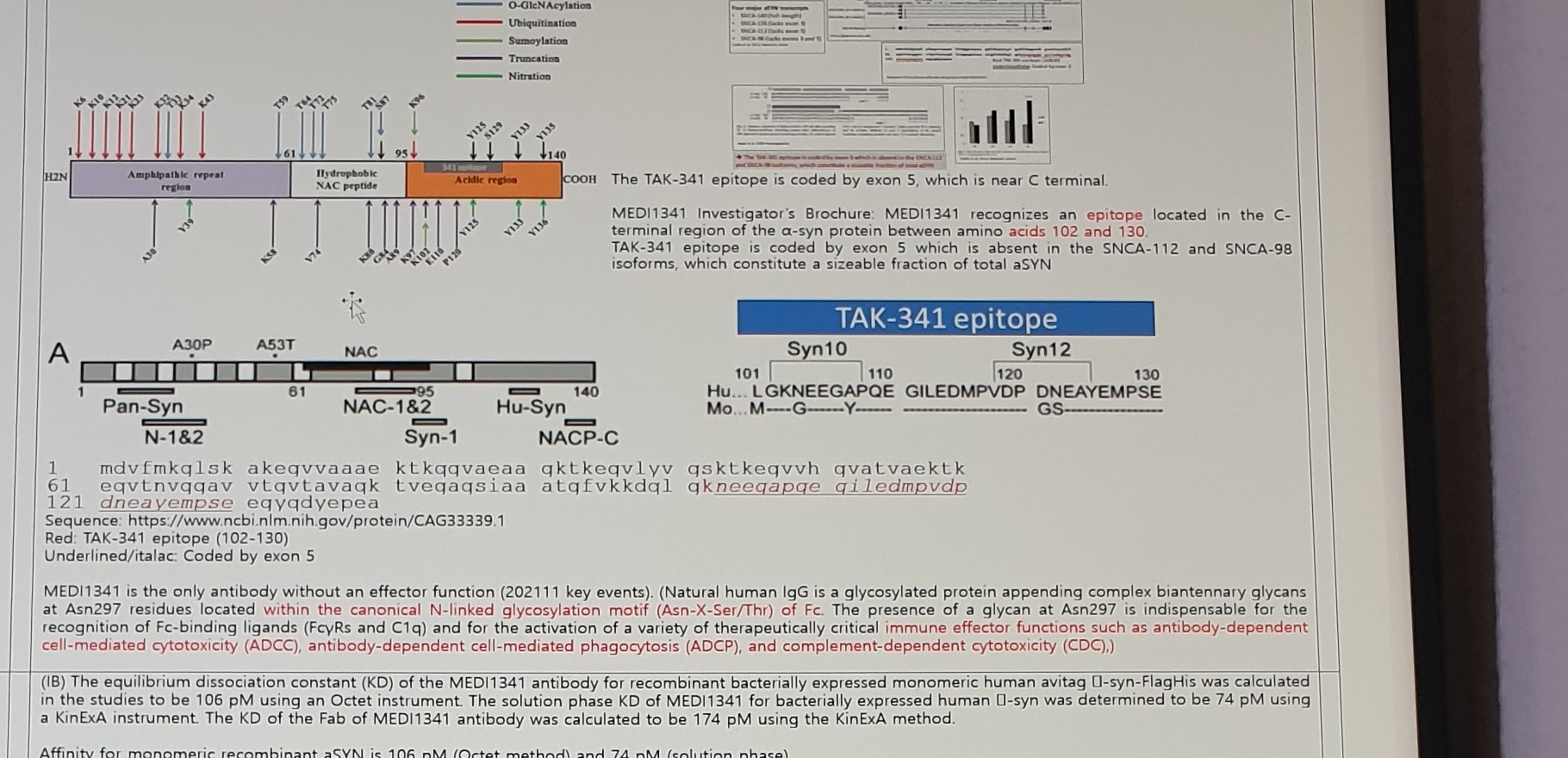
The TAK-341 epitope is coded by exon 5, which is near C terminal.MEDI1341 Investigator's Brochure: MEDI1341 recognizes an epitope located in the C-terminal region of the alpha-syn protein between amino acids 102 and 130.TAK-341 epitope is coded by exon 5 which is absent in the SNCA-112 and SNCA-98 isoforms, which constitute a sizeable fraction of total aSYN.
MEDI1341 is the only antibody without an effector function (202111 key events). (Natural human IgG is a glycosylated protein appending complex biantennary glycans at Asn297 residues located within the canonical N-linked glycosylation motif (Asn-X-Ser/Thr) of Fc. The presence of a glycan at Asn297 is indispensable for the recognition of Fc-binding ligands (FcyRs and C1q) and for the activation of a variety of therapeutically critical immune effector functions such as antibody-dependent cell-mediated cytotoxicity (ADCC), antibody-dependent cell-mediated phagocytosis (ADCP), and complement-dependent cytotoxicity (CDC).)
MEDI1341 Affinity
(IB) The equilibrium dissociation constant (KD) of the MEDI1341 antibody for recombinant bacterially expressed monomeric human avitag alpha-syn-FlagHis was calculated in the studies to be 106 pM using an Octet instrument. The solution phase KD of MEDI1341 for bacterially expressed human alpha-syn was determined to be 74 pM using a KinExA instrument. The KD of the Fab of MEDI1341 antibody was calculated to be 174 pM using the KinExA method.Affinity for monomeric recombinant aSYN is 106 pM (Octet method) and 74 pM (solution phase).Affinity for aggregated aSYN is of 37.3 - 930 pM (not substantially different than for monomeric).
Synuclein species
Ab concentration for immobilization
TAK-341 KD (M)
PRX002 KD (M)
Monomer
10 ug/mL
1.38 x 10^-10
7.38 x 10^-10
Oligomer
1 ug/mL
3.73 x 10^-11
6.23 x 10^-10
Oligomer
0.1 ug/mL
9.30 x 10^-10
1.22 x 10^-9
Figure 4
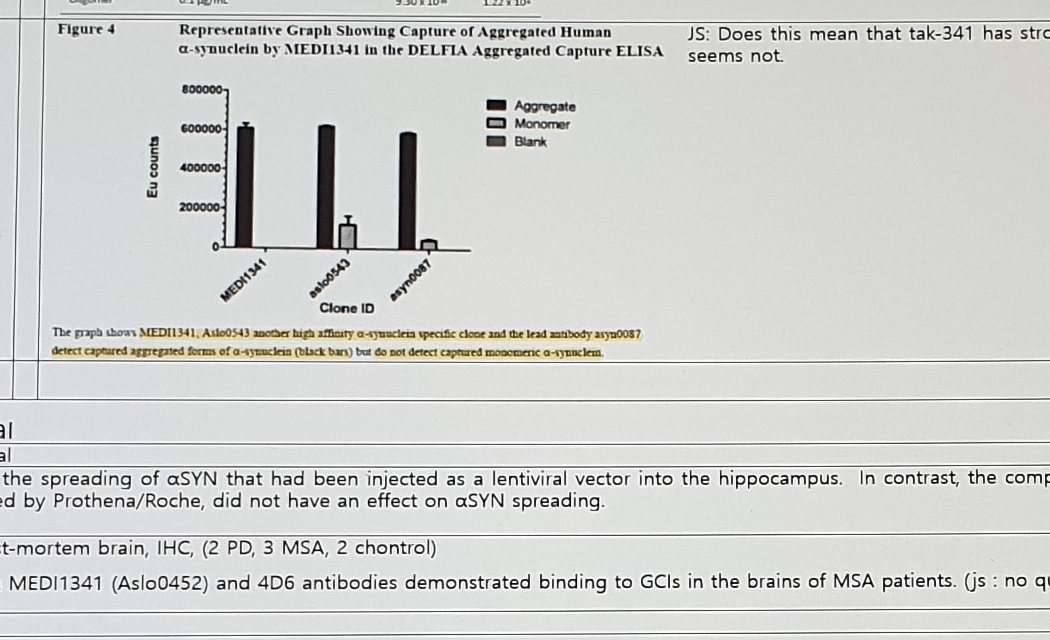
Figure 4Representative Graph Showing Capture of Aggregated Humanalpha-synuclein by MEDI1341 in the DELFIA Aggregated Capture ELISAAggregateMonomerBlankEu countsClone IDMEDI1341Aslo0543asyn0087
JS: Does this mean that tak-341 has stronger affinity to aggregated aSyn than to monomer? It seems not.
The graph shows MEDI1341, Aslo0543 another high affinity alpha-synuclein specific clone and the lead antibody asyn0087 detect captured aggregated forms of alpha-synuclein (black bars) but do not detect captured monomeric alpha-synuclein.
Preclinical
Preclinicalpreclinical
reduced the spreading of aSYN that had been injected as a lentiviral vector into the hippocampus. In contrast, the comparator antibody 9E4, which is the aSYN antibody being developed by Prothena/Roche, did not have an effect on aSYN spreading.MSA post-mortem brain, IHC, (2 PD, 3 MSA, 2 chontrol)-> both MEDI1341 (Aslo0452) and 4D6 antibodies demonstrated binding to GCIs in the brains of MSA patients. (js: no quantification was done)
PK/PD Model
PK/PD modelAbove from IB,
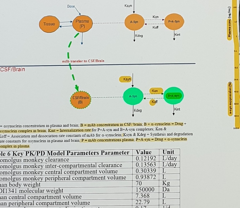
Figure 17PK/PD Model Describing the PK of MEDI1341 by a Two-Compartment Model and PD of alpha-synuclein
PlasmaTissuePlasma (P)DoseKsynA-SynKonKoffP+A-SynKdegKintmAb transfer to CSF/BrainCSF/BrainCSF/Brain (B)KsynA-SynKonKoffB+A-synKdegKint
A-syn = asynuclein concentration in plasma and brain; B = mAb concentration in CSF/brain; B + alpha-synuclein = Drug + alpha-synuclein complex in brain; Kint = Internalization rate for P+A-syn and B+A-syn complexes; Kon & Koff = Association and dissociation rate constants of mAb for alpha-synuclein; Ksyn & Kdeg = Synthesis and degradation rate constants for asynuclein in plasma and brain; P = mAb concentrations plasma; P+A-syn = Drug + alpha-synuclein complex in plasma.
Figure 18
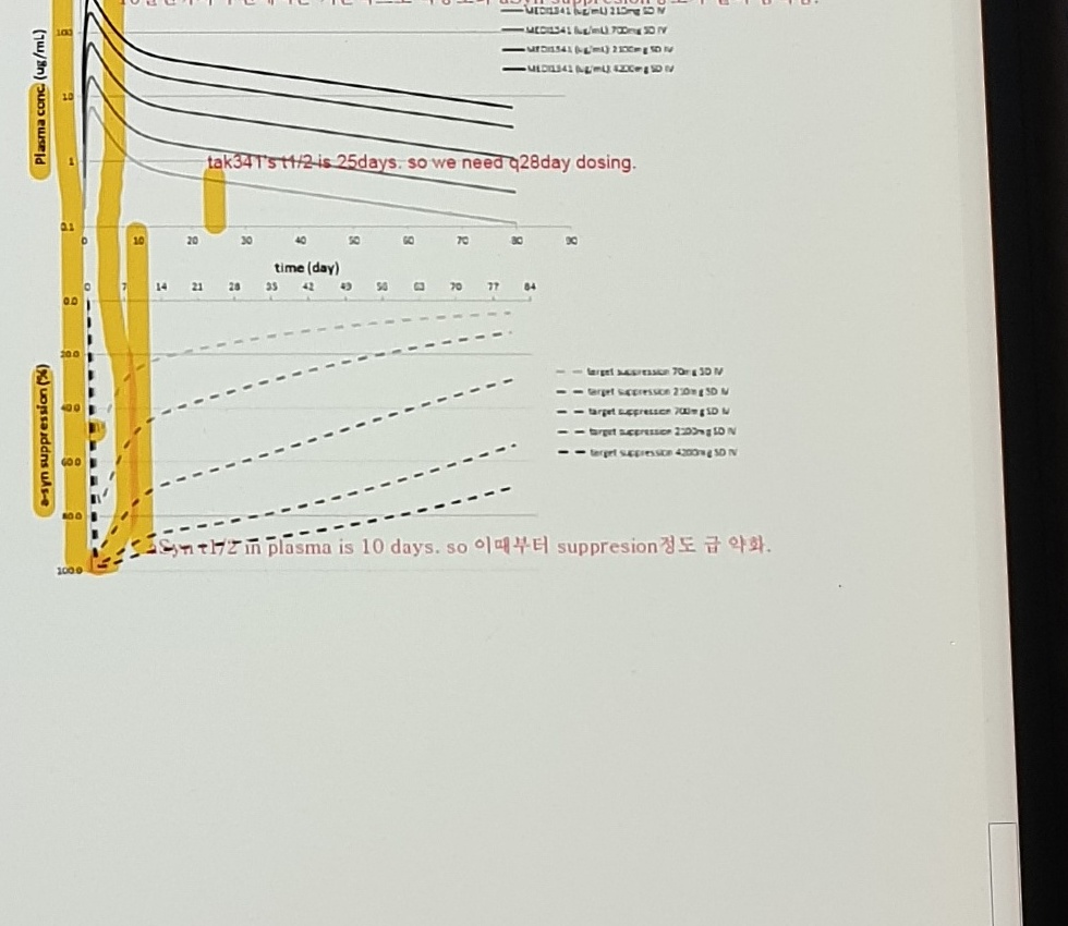
Figure 18Predicted Median Plasma Profile after Single IV Dose Administration of MEDI1341 in HumanHuman predicted plasma profiles and target suppression of MEDI1341
10일전까지 구간에서는 기본적으로 약농도와 aSyn suppression 정도가 같이 움직임.tak341's t1/2 is 25days. so we need q28day dosing.Syn t1/2 in plasma is 10 days. so 이때부터 suppression 정도 급 약화.
Table 6: Key PK/PD Model Parameters
Parameter
Value
Unit
Cynomolgus monkey clearance
0.12192
L/day
Cynomolgus monkey inter-compartmental clearance
0.13563
L/day
Cynomolgus monkey central compartment volume
0.30339
L
Cynomolgus monkey peripheral compartment volume
0.93872
L
Human body weight
70
Kg
MEDI1341 molecular weight
150000
Da
Human central compartment volume
7.368
L
Human peripheral compartment volume
22.79
L
Human central compartment elimination rate
0.17
1/d
Human clearance
1.32
L/day
Human alpha-syn elimination rate from brain
0.17
1/d
Human inter-compartmental clearance
1.46
L/day
MEDI1341 brain penetration
0.1
%
MEDI1341 binding dissociation constant
3.3
1/d
MEDI1341 affinity
0.074
nM
Human t1/2 of alpha-syn in plasma
240
h
Human t1/2 of alpha-syn in brain
240
h
Human alpha-syn basal levels in plasma
0.9
nM
Human alpha-syn basal levels in brain
0.093
nM
Table 7: Planned MEDI1341 Dose Escalations
Table 7Planned MEDI1341 Dose Escalations and Predicted % alpha-synuclein Suppression in Cerebrospinal Fluid at each Dose
Cohort
MEDI1341 Dose (mg)^a
Predicted % alpha-synuclein Suppression after Single Infusion: 28 Days after Infusion (Day 29)
Predicted % alpha-synuclein Suppression after Single Infusion: At Peak
Cohort 1
70
16
50
Cohort 5
4200
90
98
a All doses will be administered by intravenous infusion.
TxPKBrain ISF-FreePlasmaCSF
Uncertain Spans
location
text/status
reason
carryover top row
(Mazzulli et al. 2016, PMID)
PMID value is not visible in this photo.
mechanism diagram
phosphorylation / glycosylation / ubiquitination site labels
Many residue labels are too small; preserved as image asset instead of exact residue-level transcription.
sequence block
alpha-synuclein amino-acid sequence
Transcribed from visible sequence with OCR help; individual letters should be checked before using as a canonical sequence.
affinity table
KD exponents
Values are consistent with the visible pM summary, but the table exponents are tiny and should be checked against the image.
clone IDs
Aslo0543, asyn0087, Aslo0452
OCR and image distinguish l/1/o poorly.
Figure 18 legend
dose legend and target suppression legend
The right-side legend is small; doses were reconstructed from OCR plus visual evidence.
Table 7
only Cohort 1 and Cohort 5 visible
Intermediate cohort rows are not visible in this photo and likely require adjacent photo evidence.