Parkin/PARK2 Fibroblast And iPSC Mitochondrial Evidence
Zilocchi 2020 PARK2 Fibroblast Continuation
Continuation note:
(Zilocchi, 2020 #901) n=5, 개별 환자자료유, genotyping data 있음. Age: 15-75. Proteomics, ↓ MMP (fig2c, dmso상황에서 이미 낮고 CCCP로 추가 감소 없음. 나이 다양한데도 별로 variance 없이 감소되어 있음.), = mito network morphology (authors: While DRP1 localizes to depolarized mitochondria, OPA1 and MFN1 levels are not altered. As a whole, it seems that even if fission is triggered (DRP1 accumulation), fusion is not blocked (lack of OPA1 and MFN1 elimination). No PINK1 accumulation (아예 not detectible at baseline) CCCP treatment determined the accumulation of PINK1 in control subjects. On the other hand, PINK1 levels were drastically reduced in skin fibroblasts from PARK2-mutated patients exposed to CCCP,Patient-table anchors:
| Subject | Age at onset | Age at skin biopsy | PARK2 mutations |
|---|---|---|---|
| P1 | 60-65 | 70-75 | Del_1/p.R275W |
| P2 | 15-20 | 40-45 | Del_3-4-5/p.R33X |
| P3 | 10-15 | 15-20 | Dup_2/Del_3-4-5 |
| P4 | 40-45 | 50-55 | p.Q34Rfs x X5 homo |
| P5 | 20-25 | 30-35 | p.Thr240Met/Del3 |
| C1-C5 | CTRL | 25-30, 25-30, 30-35, 50-55, 50-55 | CTRL |
Figure caption anchors:
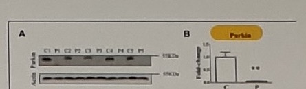
FIGURE 1 | Parkin levels are reduced in PARK2-mutated patients.
(A) Representative western blot image of Parkin protein in primary skin fibroblasts of PARK2-mutated patients (P1, P2, P3, P4, and P5) and control subjects (C1, C2, C3, C4, and C5). (B) Relative quantification expressed as mean ± SEM (three technical replicates). Data were analyzed by two-tailed, unpaired Student's t-test by comparison of PD patients and control subjects. **p < 0.001.
Zilocchi Mitochondrial Proteomics And PhRET Proposal
[mitochondrial Proteomics:]
- mitochondrial fraction:
  - 649 proteins identified,
  - NDUFS8, NDUFS7 (PSST) 없네
  - 227 significantly altered (↑ NDUFV1, ↓ (control군에서만 발견됨) NDUFS3) -> 227만 가지고 network-based analysis (PPI) 시행: UPR (several HSPs) and small GTPases (Rab proteins, Rab7A)-mediated signal transduction as the two main pathways altered.

- total fraction:
  - 1457 proteins identified,
  - ndu 거의 없네 (only two ...)
  - 168 significantly altered

[PhreT proposal]
- mito fraction vs total fraction
- how can we ensure to include MC1 PET-specific proteins in the proteomics?
Koentjoro Mitophagy And Rescue Notes
(Koentjoro, 2017 #711) fig 3e,
nix 없는 상태에서, CCCP 줄 때,, mtDNA contect 감소가 parkin-pd fibroblast 에서는 없네 (ie mitophagy 안 되네)
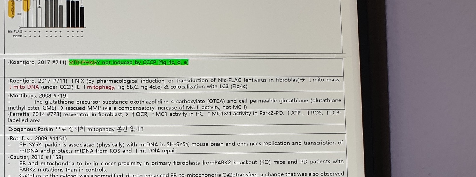
(Koentjoro, 2017 #711) MITOPHAGY not induced by CCCP (fig 4c, d, e)
Correction
(Koentjoro, 2017 #711) ↑ NIX (by pharmacological induction, or Transduction of Nix-FLAG lentivirus in fibroblas) -> ↓ mito mass, ↓ mito DNA (under CCCP, IE ↑ mitophagy, Fig 5B,C, fig 4d,e) & colocalization with LC3 (Fig4c)
(Mortiboys, 2008 #719)
- the glutathione precursor substance oxothiazolidine 4-carboxylate (OTCA) and cell permeable glutathione (glutathione methyl ester, GME) -> rescued MMP (via a compensatory increase of MC II activity, not MC I)
(Ferretta, 2014 #723) resveratrol in fibroblast, -> ↑ OCR, ↑ MC1 activity in HC, ↑ MC1&4 activity in Park2-PD, ↑ ATP, ↓ ROS, ↑ LC3-labelled area
Exogenous Parkin 으로 정확히 mitophagy 본건 없네?mtDNA, ER-Mitochondria, And Fibroblast Rescue Literature
(Rothfuss, 2009 #1151)
- SH-SY5Y: parkin is associated (physically) with mtDNA in SH-SY5Y, mouse brain and enhances replication and transcription of mtDNA and protects mtDNA from ROS and ↑ mt DNA repair

(Gautier, 2016 #1153)
- ER and mitochondria to be in closer proximity in primary fibroblasts from PARK2 knockout (KO) mice and PD patients with PARK2 mutations than in controls.
- Ca2+ flux to the cytosol was also modified, due to enhanced ER-to-mitochondria Ca2+ transfers, a change that was also observed in neurons derived from induced pluripotent stem cells of a patient with PARK2 mutations.
- the abundance of the Parkin substrate mitofusin 2 (Mfn2), which is known to modulate the ER-mitochondria interface, to be specifically higher in the mitochondrion-associated ER membrane compartment in PARK2 KO tissue.
- the exogenous expression of normal Parkin restored cytosolic Ca2+ transients in fibroblasts from patients with PARK2 mutations.

(Hsieh, 2016 #1154) fibroblast Parkin255A EX3-4Del

(Haylett, 2016 #1155) n=3, fibroblast. Deletion exon 3-4 hom, and two Deletion exon 4 hom
- ↑ mitochondrial respiratory rates (Seahorse flux analysis): ↑ Oxygen consumption (basal, devoted to ATP synthesis and due to the natural proton leak across the inner mt membrane)
- fragmented mitochondrial networks (↓ branching, = elongation)
- = MMP
- ↑ cell growth of the parkin-mutant fibroblasts
Choong 2020 evidence is preserved separately because the figure panels and interpretation are compact and partly watermarked.
Choong, 2020 #1193) ↑ TOM40, TOM20
여기서, fibroblast 에서 TOM20 증가한 것이 '↑ Extracellular mito'라는데, 아마 media 로 WB 한거라네 (p18)
Fibroblast, Lipidomics, And General Translation Notes
(Imaizumi, 2012 #1328) In PARK2 iPSC-derived neurons, but not PARK2 fibroblasts or iPSCs, abnormal mitochondrial morphology (highly electron-dense matrix and swollen mitochondrial cristae within the inner mitochondrial membrane (IMM)(Figure 3A, black arrowheads).
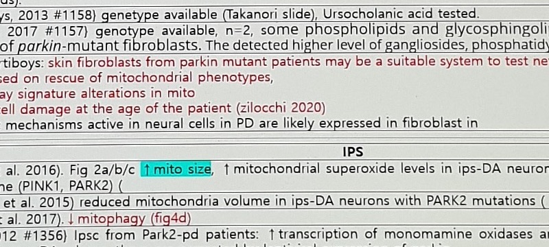
(Mortiboys, 2013 #1158) genotype available (Takanori slide), Ursocholanic acid tested.
(Lobasso, 2017 #1157) genotype available, n=2, some phospholipids and glycosphingolipids were altered in the lipid profiles of parkin-mutant fibroblasts. The detected higher level of gangliosides, phosphatidylinositol, and phosphatidylserine
2008 Mortiboys: skin fibroblasts from parkin mutant patients may be a suitable system to test new therapeutic approaches, at least those based on rescue of mitochondrial phenotypes,
- display signature alterations in mito
- the cell damage at the age of the patient (zilocchi 2020)
- basic mechanisms active in neural cells in PD are likely expressed in fibroblast inLeft-side comparison note:
Matching Control feasible? : doable, but the same passage number, doubling time, culture condition matters will be challenging.Comparative postmortem note:
Cf) (Grünewald, 2016 #935) sPD (parkin-PD 아님!) postmortem brain,
Whereas mitochondrial mass was unchanged in single SN neurons from IPD patients, we observed a significant reduction in the abundances of CI and II subunits in dopaminergic neurons
- fewer transcription/replication-associated mtDNA molecules and an overall reduction in mtDNA copy number

some proliferator-activated receptor-gamma coactivator-1α (PGC-1α): master regulator of mitochondrial functions and oxidative metabolism
iPSC Evidence Summary
Left-side mutation anchor:
iPSC
c.1072Tdel
Homozygous
p.A324 fsX110| Study | Evidence summary |
|---|---|
| Chung et al. 2016 | Fig 2a/b/c; ↑ mito size; ↑ mitochondrial superoxide levels in ips-DA neurons from patients with mutations in either gene (PINK1, PARK2) |
| Shaltouki et al. 2015 | reduced mitochondria volume in ips-DA neurons with PARK2 mutations |
| Suzuki et al. 2017 | ↓ mitophagy (fig4d) |
| Jiang, 2012 #1356 | Ipsc from Park2-pd patients: ↑ transcription of monomamine oxidases and oxidative stress, ↓ DA uptake & ↑ spontaneous DA release, these are corrected by lentiviral expression of parkin |
| Okarmus, 2020 #1331 | isogenic Ips. proteomic analysis: 119 proteins altered. Swollen, mito with ↓ matrix density and irregular cristae, increased TOM20 (Fig4b) as earlier reported29 |
| Bogetofte, 2019 #1332 | PARK2 KO did not significantly affect the total number of mitochondria as measured by TOM20 immunofluorescence staining (Figures 4A,B). However, the PARK2 KO neurons contained a significantly increased area of TOM20 immunoreactivity, indicating an overall larger mitochondrial area per cell (Figures 4A,C) |
| Kumar, 2020 #1333 | ↓ OCR, ↓ MMP, ↓ TOM20 (fig7n) |
| Rakovic, 2015 #1334 | review |
Translation Plan Fibroblast And Leukocyte Notes
Translation plan anchors:
| Question / phase | Timing | Need / comment | Evidence |
|---|---|---|---|
Fibroblasts from Parkin-PD show mitophagy? | blank | 대표적 흔한것들에 필요 | Papers exist (Koentjoro, 2017 #711) |
Corrected by exogenous Parkin? | Before clinical trial | 대표적 흔한것들에 필요 | No evidence but T. Nishimura in NS-DDU PhRET showed this in PARK2 KO iPSC-DA neurons (is this patient-derived?) |
| blank | Clinical trial | blank | blank |
Leukocyte/WBC note:
(Müftüoglu, 2004 #1027) 10 patients from 6 families with parkin gene mutations, 20 patients with idiopathic PD, ...

Method: venous blood samples collected -> WBC isolated -> mito isolated (fractionation) 즉 culture 아님!

Results: ↓ MC I and IV activities in leukocytes from patients with idiopathic PD, and decreased complex I activity, with normality of complex IV, in patients with parkin mutations (by 62.5%) and Spd (by 64.5%): table 2, fig1 (seems clearly differentiated)
Uncertain Spans
- Lower leukocyte figure/table is only partially visible; numerical values and group distinctions are uncertain.