pS65-Ub Postmortem Evidence, NBB Comparison, And Assay Development Notes
Hou 2018 p-S65-Ub Aging And LBD Figures
Source/context note:
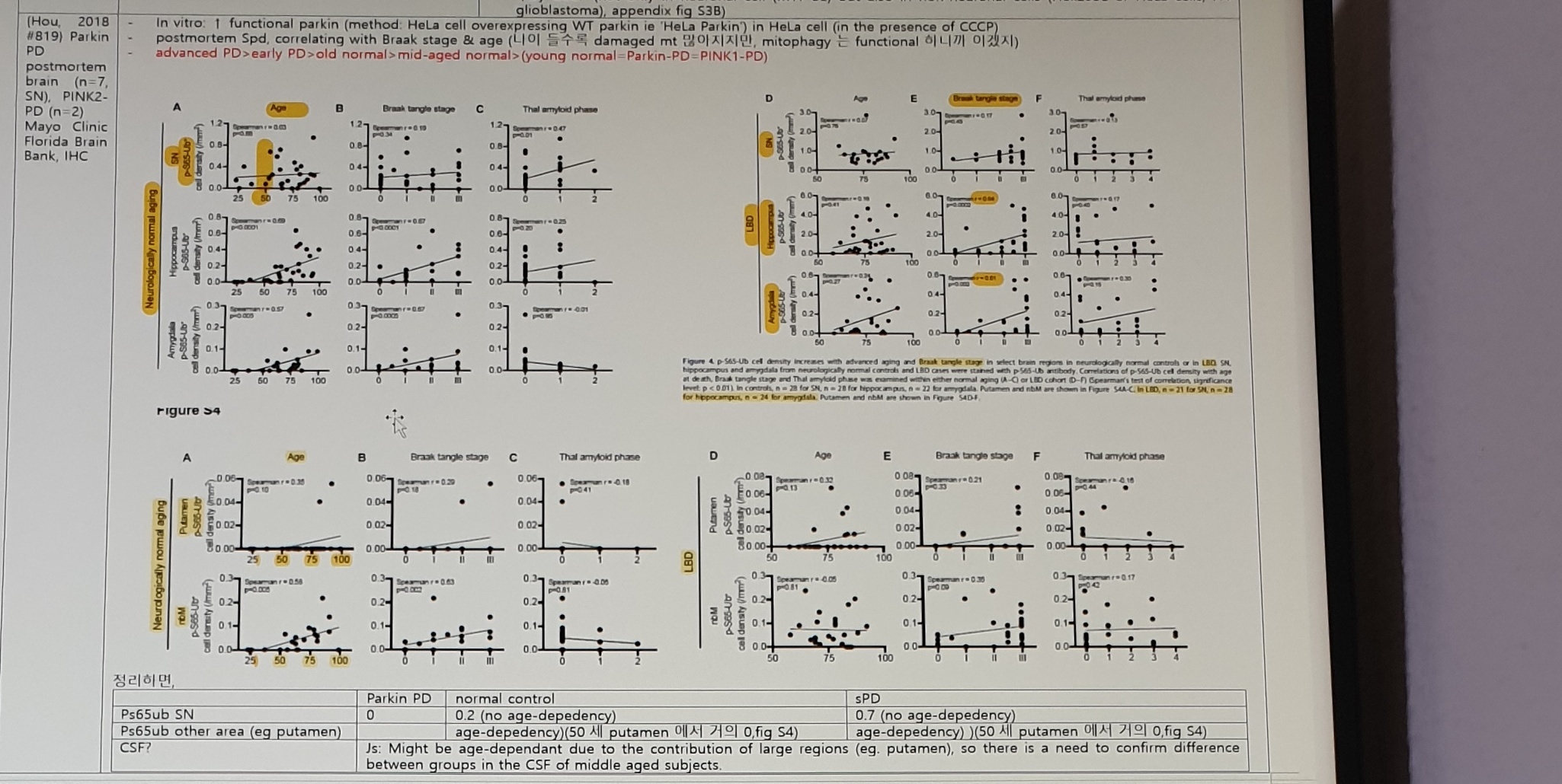
(Hou, 2018 #819) Parkin PD postmortem brain (n=7, SN), PINK2-PD (n=2)
Mayo Clinic Florida Brain Bank, IHC

In vitro: ↑ functional parkin
(method: HeLa cell overexpressing WT parkin ie 'HeLa Parkin')
in HeLa cell (in the presence of CCCP)

postmortem sPD, correlating with Braak stage & age
(나이 들수록 damaged mt 많아지지만, mitophagy는 functional 하니까 이겠지)

advanced PD > early PD > old normal > mid-aged normal >
(young normal = Parkin-PD = PINK1-PD)
Figure labels:
Figure S4
Neurologically normal aging
LBD
SN p-S65-Ub cell density
Hippocampus p-S65-Ub cell density
Amygdala p-S65-Ub cell density
Putamen p-S65-Ub cell density
nbM p-S65-Ub cell density
Age
Braak tangle stage
Thal amyloid phase
Caption fragment near the upper-right figure:
Figure 4. p-S65-Ub cell density increases with advanced aging and Braak
tangle stage in select brain regions in neurologically normal controls or
in LBD.
Summary Table
| Marker / question | Parkin PD | normal control | sPD |
|---|---|---|---|
Ps65ub SN | 0 | 0.2 (no age-depedency) | 0.7 (no age-depedency) |
Ps65ub other area (eg putamen) | age-depedency)(50 세 putamen 에서 거의 0, fig S4) | age-depedency)(50 세 putamen 에서 거의 0, fig S4) | |
CSF? | Js: Might be age-dependant due to the contribution of large regions (eg. putamen), so there is a need to confirm difference between groups in the CSF of middle aged subjects. |
Hou 2018 Reduced Or Absent p-S65-Ub Staining Figure
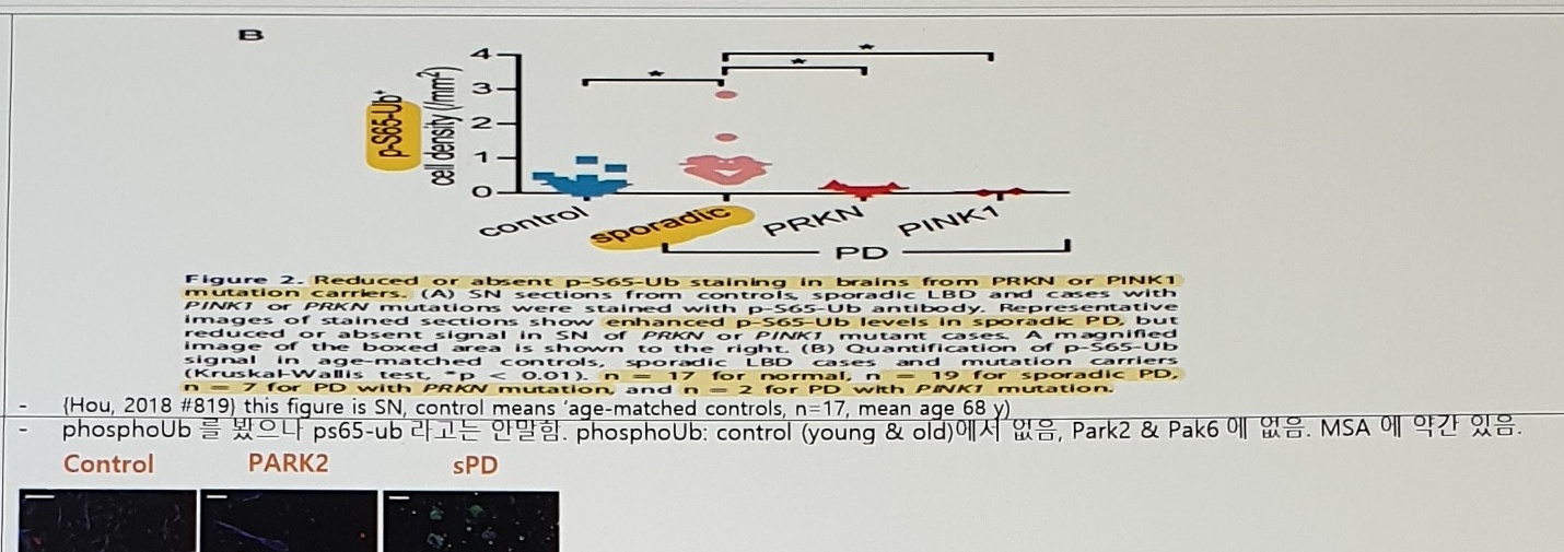
Caption text:
Figure 2. Reduced or absent p-S65-Ub staining in brains from PRKN or PINK1
mutation carriers.
Figure/caption anchors:
control
sporadic
PRKN
PINK1
PD
p-S65-Ub cell density

SN sections from controls, sporadic LBD and cases with PINK1 or PRKN
mutations were stained with p-S65-Ub antibody.

Representative images of stained sections show enhanced p-S65-Ub levels in
sporadic PD, but reduced or absent signal in SN of PRKN or PINK1 mutant cases.

(B) Quantification of p-S65-Ub signal in age-matched controls, sporadic LBD
cases and mutation carriers.
Note beneath the figure:
(Hou, 2018 #819) this figure is SN, control means 'age-matched controls',
n=17, mean age 68 y
Shiba-Fukushima 2017 Phospho-Ub IHC Note
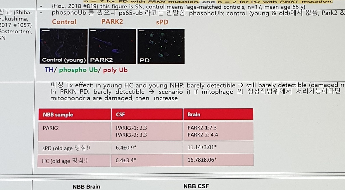
Source/context note:
참고: (Shiba-Fukushima, 2017 #1057) Postmortem, SN
phosphoUb 를 봤으나 ps65-ub 라고는 안말함.
phosphoUb: control (young & old)에서 없음, Park2 & Pak6 에 없음.
MSA 에 약간 있음.
Image labels:
Control
PARK2
sPD
Control (young)
PARK2
PD
TH / phospho Ub / poly Ub
Expected Treatment Effect And PRKN-PD Scenario Notes
예상 Tx effect in young HC and young NHP:
barely detectible -> still barely detectible
(damaged mitochondria 적으므로)

In PRKN-PD: barely detectible -> scenario
i) if mitophagy 의 정상적범위에서 처리가능하다면 no increase,
ii) if mitophagy 급증 혹은 여전히 mitochondria are damaged, then increase
NBB Sample Brain / CSF Values
| NBB sample | CSF | Brain |
|---|---|---|
PARK2 | PARK2-1: 2.3PARK2-2: 3.3 | PARK2-1: 7.3PARK2-2: 4.4 |
sPD (old age 명심!) | 6.4±0.9* | 11.14±3.01* |
HC (old age 명심!) | 6.4±3.4* | 16.78±8.06* |
NBB Brain / CSF pS65Ub K48 Plots
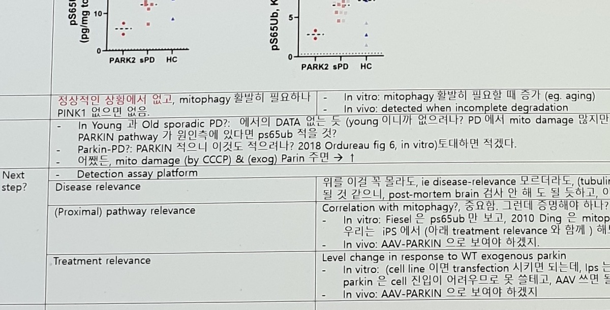
Plot labels:
NBB Brain
pS65Ub, K48 (pg/mg total protein)
PARK2
sPD
HC

NBB CSF
pS65Ub, K48 (pg/mL)
PARK2
sPD
HC
Interpretation Notes Before Next-Step Plan
정상적인 상황에서 없고, mitophagy 활발히 필요하나
PINK1 없으면 없음.

In vitro: mitophagy 활발히 필요할 때 증가 (eg. aging)
In vivo: detected when incomplete degradation

In Young 과 Old sporadic PD?: 에서의 DATA 없는 듯
(young 이니까 없으려나? PD 에서 mito damage 많지만 mitophagy 는 잘되고 있다면 ps65ub 많을 것, PINK1-)

PARKIN pathway 가 원인측에 있다면 ps65ub 적을 것?

Parkin-PD?: PARKIN 적으니 이것도 적으려나?
2018 Ordureau fig 6, in vitro) 토대하면 적겠다.

어쨌든, mito damage (by CCCP) & (exog) Parin 주면 -> ↑
Next-Step Development Plan
| Area | Plan text |
|---|---|
| Detection assay platform | Detection assay platform |
| Disease relevance | 위를 이걸 꼭 몰라도, ie disease-relevance 모르더라도, (tubulin acetylation 경우처럼) proximal PD marker 로 사용하면 될 것 같으니, post-mortem brain 검사 안 해도 될 듯하고, 이건 안 중요하지 않나? |
| (Proximal) pathway relevance | Correlation with mitophagy?, 중요함. 그런데 증명해야 하나?- In vitro: Fiesel 은 ps65ub 만 보고, 2010 Ding 은 mitophagy 만 봤으니, 둘을 한꺼번에 본 것이 없는 듯 한데?우리는 iPS 에서 (아래 treatment relevance 와 함께) 해보자.- In vivo: AAV-PARKIN 으로 보여야 하겠지. |
| Treatment relevance | Level change in response to WT exogenous parkin- In vitro: (cell line 이면 transfection 시키면 되는데, Ips 는 어떻게 exogenous parkin 을 존재시키나? Recombinant parkin 은 cell 진입이 어려우므로 못 쓸테고, AAV 쓰면 될 것임.- In vivo: AAV-PARKIN 으로 보여야 하겠지 |
Correction Of pS65-Ub / External Assay Notes
Correction Of pS65-Ub
| Readout / model | Source | No CCCP, No parqin | CCCP only | Exogenous Parkin only | CCCP + Exogenous Parkin |
|---|---|---|---|---|---|
pS65-Ub; HeLa cell | (Fiesel, 2015 #700) fig2a | Not detectible | ↑ | Not detectible | ↑↑ |
mitophagy; HeLa cell | 2010 Ding | A little | ↑ (fig 1) | No increase | ↑ |
HCT116 cells | 2010 Ding | ↑ | |||
MEF cells | 2010 Ding | ↑ | |||
Fibroblast from Parkin-PD | (Koentjoro, 2017 #711) | No change 인 듯 (fig 4c) | |||
Fibroblast from Parkin homoz mutation but no PD | (Koentjoro, 2017 #711) | To be checked! |
External Assay Notes
Ub assays (external)
Takeda

Capture Ab
Ubiquitin
mouse
mono, P4D1 (CST, 3936S),

Detection Ab
Phospho-ub
(K63 pUb tetra?)
rabbit poly

Standard protein -
K63 pUb tetra
Uncertain Spans
PINK2-PD (n=2)in the Hou 2018 source note conflicts with nearbyPINK1figure labels.- Hou 2018 figure panel labels, axis values, Spearman
r, p-values, and sample counts are image-primary; use the embedded figures before extracting exact numeric values. - NBB table values, plus/minus values, asterisks, group labels, and axis units are small and uncertain in the photo.
- Column/row alignment in the correction table is uncertain because the table is partially cropped at the lower edge and photographed at an angle.